Global view of human protein glycosylation pathways and functions
Authors: Katrine T. Schjoldager, Yoshiki Narimatsu, Hiren J. Joshi & Henrik Clausen
Abstract: Glycosylation is the most abundant and diverse form of post-translational modification of proteins that is common to all eukaryotic cells. Enzymatic glycosylation of proteins involves a complex metabolic network and different types of glycosylation pathways that orchestrate enormous amplification of the proteome in producing diversity of proteoforms and its biological functions. The tremendous structural diversity of glycans attached to proteins poses analytical challenges that limit exploration of specific functions of glycosylation. Major advances in quantitative transcriptomics, proteomics and nuclease-based gene editing are now opening new global ways to explore protein glycosylation through analysing and targeting enzymes involved in glycosylation processes. In silico models predicting cellular glycosylation capacities and glycosylation outcomes are emerging, and refined maps of the glycosylation pathways facilitate genetic approaches to address functions of the vast glycoproteome. These approaches apply commonly available cell biology tools, and we predict that use of (single-cell) transcriptomics, genetic screens, genetic engineering of cellular glycosylation capacities and custom design of glycoprotein therapeutics are advancements that will ignite wider integration of glycosylation in general cell biology
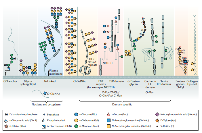
Figure 1| Main classes of glycoconjugates of the human cellular glycome. Depiction of the key components of the cellular glycome, highlighting types of glycosylation that are specific to distinct protein classes or protein domains. The glycans depicted are only illustrative examples of the glycan structures that can be synthesized by the different types of glycosylation pathways. N-glycans and most GalNAc-type O-glycans are widely found on most proteins trafficking the cellular secretory pathway, whereas the occurrence of domain-specific glycans is limited to specific protein domains. The enzymatic processes that orchestrate glycosylation of the different types of glycoconjugates are partly distinct and partly overlapping. The initial attachment of the first monosaccharide (or oligosaccharide for N-glycosylation) to proteins represents the key initiation step that determines which proteins and positions become glycosylated. These initiation steps are distinct for the different types of glycosylation pathways and, to large extent, direct the structures of glycans that are generated. Some overlap in the later processing steps that involve elongation, branching and capping of oligosaccharides is found among N-glycosylation and several types of O-glycosylation as well as in glycosphingolipid biosynthesis (Fig. 3). The background colour scheme is organized according to the colour of the first monosaccharide attached to the core protein, except for glycosylphosphatidylinositol (GPI)-anchored proteins and glycolipids (shown in grey). The colouring scheme is useful for distinguishing the protein glycosylation pathways involved in the synthesis of different types of glycans215 (see also Table 1). Glycan symbols are drawn according to the Symbol Nomenclature for Glycans (SNFG) format246. Sugar repeat units are indicated by square brackets with ‘n’ to indicate a number of possible repeats. EC, extracellular cadherin; EGF, epidermal growth factor; Hyl, hydroxylysine; IPT, immunoglobulin-like, plexin, transcription factor; LDLR, low-density lipoprotein receptor; NS, non-specific; TSR domain, thrombospondin type 1 repeat domain.